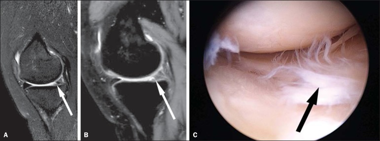
Figure 1.
A 36-year-old woman with a tear of the medial meniscus. Sagittal MRI (A: 2D TSE; and B: 3D TSE volume isotropic turbo spin-echo acquisition [VISTA]) of the knee depicting a tear of the posterior horn of the medial meniscus (arrows). C: Corresponding arthroscopic correlation of the tear (arrow).

