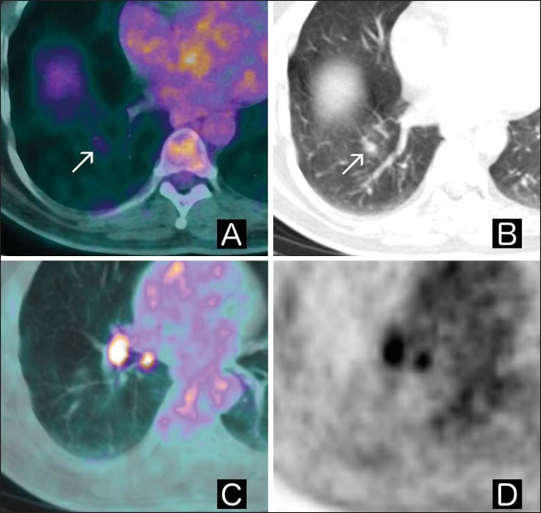
Figure 3.
A: Axial fused 18F-FDG PET/CT image. B: Axial chest CT image. C: Axial fused 18F-FDG PET/CT image. D: PET axial image. Patient with a history of gastrointestinal stromal tumor presenting with indeterminate pulmonary nodule in the right lower lobe in a previous CT scan. Upon 18F-FDG PET/CT examination, the nodule displayed slightly increased glycolytic metabolism when compared with normal lung parenchyma (arrows in A and B), together with hypermetabolic ipsilateral hilar lymph nodes (C and D). The patient underwent surgery, which further confirmed the diagnosis of tuberculosis.

