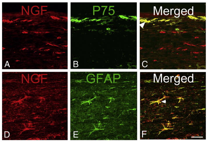
Fig. 2.
Photomicrographs of NGF immunoreactive cells in longitudinal sections of the T5 spinal cord at 7 days after T4 spinal cord injury. A–C) NGF is expressed in cells at and below the pial surface of the cord (A) and is co-localized with the low affinity neurotrophin receptor p75NTR (B) in cells adjacent to the pia that may be leptomeningeal cells (C, arrow head). D–F) NGF is expressed in large stellate cells in the white matter (D) that are immunoreactive for glial acidic fibrillary protein (E, GFAP), identifying them as astrocytes (F, arrow head). Scale bar in F is 50 μm and refers to all panels.
Adapted, with permission, from Brown et al. (2004).

