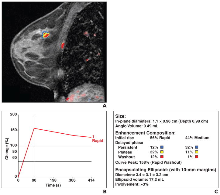
Fig. 1. 51-year-old woman with grade 2 estrogen receptor–positive, progesterone receptor–positive, and ERRB2-negative invasive ductal carcinoma.
A, Sagittal contrast-enhanced, T1-weighted volume imaging for breast assessment (VIBRANT, GE Healthcare) fat-suppressed sequence shows computer-assisted detection color overlay over breast mass. Areas in red indicate rapid washout type delayed enhancement pattern, areas in yellow indicate plateau type delayed enhancement pattern, and areas in blue indicate persistent type delayed enhancement pattern. B, Graph of worst kinetics pattern within tumor shows rapid initial enhancement and rapid washout type curve. C, Volumetric assessment by computer-assisted detection calculates volume of tumor and enhancement composition of tumor mass. With respect to initial enhancement phase, 56% of tumor shows rapid wash-in and 44% shows medium rate of wash-in. For delayed enhancement component, only 13% (12% + 1%) of mass showed rapid washout type pattern; 44% (12% + 32%) of mass contained delayed persistent type curve, and 43% (32% + 11%) contained plateau type curve.

