
Figure 5. Brain activation by visual food cues pre- and post-standardized meal in low and high anxiety women.
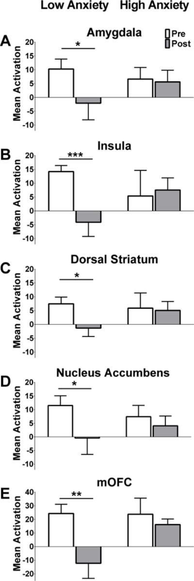
Among low anxiety women, brain activation by fattening vs. non-fattening food cues was significantly suppressed by a standardized meal in each of the five ROIs (A–E bars on left, Nucleus Accumbens, Amygdala, Insula, Dorsal Striatum, and mOFC). Women with high anxiety did not show meal-induced changes in brain activation in any of the ROIs (A–E bars on right). Data are mean parameter estimates for each ROI ± SEM pre- (white bars) and post- (gray bars) standardized meal between low and high anxiety women (left bars and right bars, respectively). Bilateral regions (A–D) were averaged. P-values derived from generalized estimating equations. *P<0.050, **P<0.010, ***P<0.001 pre-meal vs. post-meal in low anxiety women. Data are adjusted for BMI.