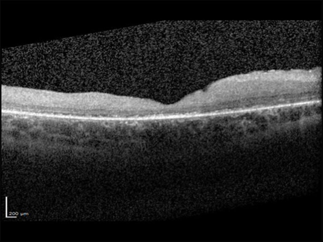
Figure 1.

Spectral domain optical coherence tomograph (SD-OCT) of the macula of the right eye showing irregular foveal contour, epiretinal membrane, and retinal thinning with subfoveal attenuation and loss of outer retinal laminar structures that include the external limiting membrane, ellipsoid zone and interdigitation zone.
