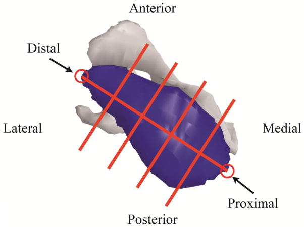
Figure 3.
A supraspinatus divided into five sections. A line connecting the most proximal and most distal points was then divided into 5 equal length segments. Planes perpendicular to the line at the ends of each segment were used to separate the muscle into 5 equal length sections. Voxels were assigned to each section based on their locations relative to the dividing planes. Using this same approach, infraspinatus and subscapularis were divided into five sections, while teres minor was divided into three sections, due to its small size.

