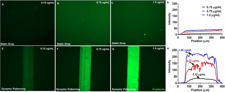
FIG. 2.
Immunofluorescence images of glass coverslips coated with mP-selectin via static drop and μFP. Immunofluorescent images of glass coverslips coated with different concentrations of mP-selectin using either a standard static drop procedure ((a)–(c)) or the μFP technique ((e)–(g)). Fluorescence intensity line scans of the static drop (d) and the μFP (h) coated substrates revealed a 5-fold higher amount of protein deposited by the μFP method. The protein line patterns were formed by perfusing the different mP-selectin solutions through microfluidic channels (300 μm × 50 μm cross-section) at a perfusion shear stress of 16.0 dyn/cm2 for 15 min.

