Fig. 2.
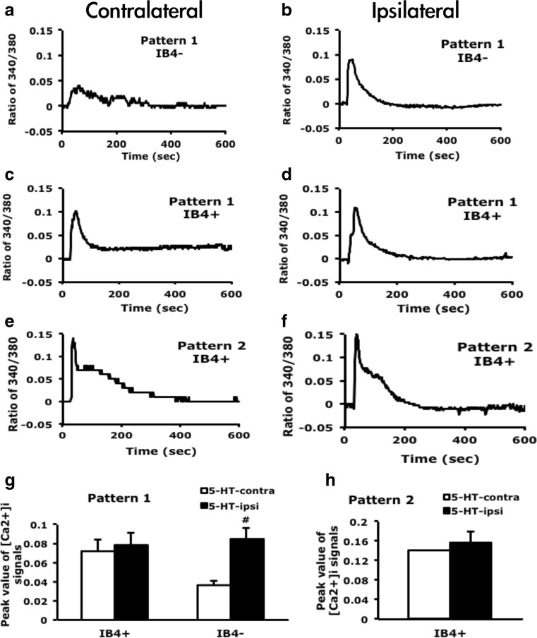
IB4-negative neurons show enhanced calcium signals after 5-HT injection. Lumbar 4–6 dorsal root ganglia (DRG) ipsilateral or contralateral to the 5-HT-injected mouse paw were cultured for 12 h, then stimulated with 5-HT (1 μM), and intracellular calcium change was recorded for 600 s. After recording, neurons were pulse labeled with IB4-FITC (5 μg/ml) for 10 min. Time-dependent mean calcium increase in IB4-negative (IB 4 −) neurons (a, b) and IB4-positive (IB 4 +) neurons (c–f). The peak values were represented in the histograms (g, h). Comparisons between ipsilateral and contralateral DRG neurons of 5-HT-injected animals were done by two-way ANOVA with a post hoc Bonferroni test. # p < 0.05

