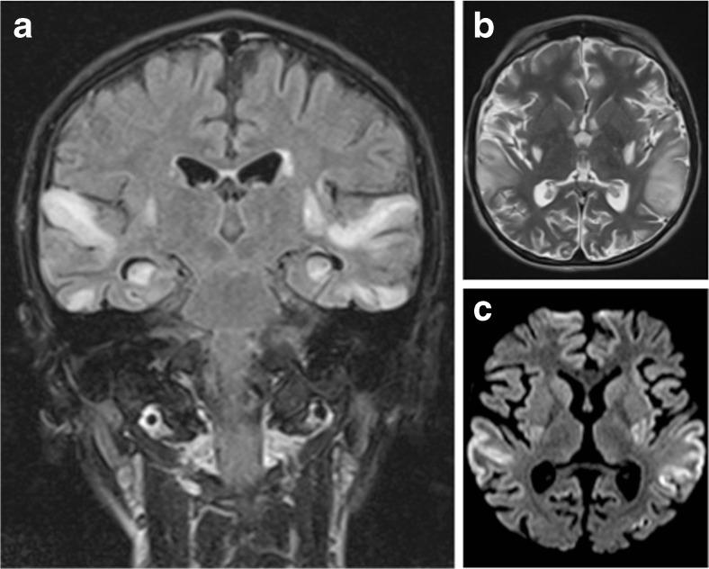
Fig. 1.
Brain MRI scan 72 h after onset of symptoms. a Coronal FLAIR image shows symmetrical high signal intensities lesions in multiple arterial territories. b Axial T2-weighted image demonstrating high signal cortical and subcortical lesions bilaterally in the edematous superior temporal gyri. c Axial diffusion-weighted image (DWI) demonstrating high signal areas in the same regions

