
Figure 1. Western blotting analysis of p22phox expression in mouse eye.
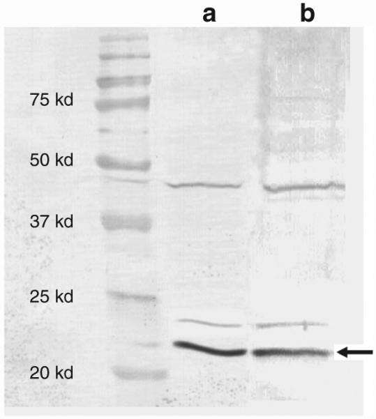
p22phox expression was detected using western blotting in (a) retinal as well as (b) retinal pigment epithelial (RPE) extracts. Protein extracts from retina and RPE/choroid were isolated by sonication, separated on 12% polyacrylamide gels, and transferred to a nitrocellulose membrane, which was then treated with a polyclonal antibody to p22phox. Antibody antigen complexes were detected using alkaline phosphatase-conjugated anti-rabbit immunoglobulin G antibody, and the filter was developed using NBT/BCIP.