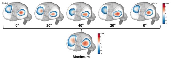
Figure 2. Dynamic cartilage contact.
Tibiofemoral kinematics were used to characterize regions of proximity between the tibial and femoral cartilage. Contact maps for each subject were created by identifying the greatest proximity of each face of the cartilage mesh over a flexion-extension motion cycle.

