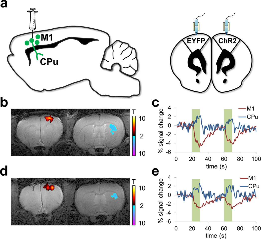
Figure 2. Study design: Cohort 2.
(a) Schematic of ofMRI experimental paradigm. Rats were deeply anesthetized with isoflurane (2%), and the primary motor cortex was targeted for optogenetics. To preferentially target cortical pyramidal cells, an adeno-associated virus was used carrying the gene encoding channelrhodopsin-2 (ChR2) fused to an enhanced yellow fluorescent protein (EYFP) or only EYFP. Chronically implanted optic fibers were placed above viral infusion sites. (b)–(c), (d)–(e): Representative t-scored functional activation maps from two rats, overlaid on same-subject T2-weighted anatomical images (b, d). Corresponding time-courses of CBV changes in motor cortex and dorsolateral striatum (c, e).

