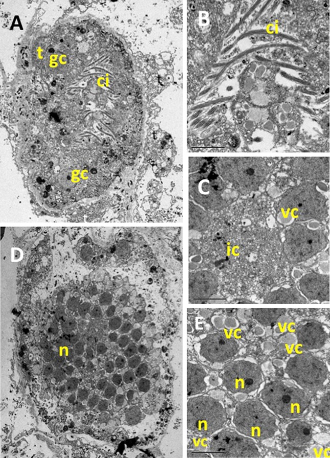
Fig. 7.

TEM pictures of a P. magna early larvae (stomoblastula). a Section surrounded by maternal trophocytes (t) showing macromeres and nucleolate micromeres and the two granular nucleolate cells (gc) that will move to the internal space of the amphiblastula. b Detail of the inner zone showing the cilia of the epithelial layer already differentiated but still inwards. c Detail of the anterior zone of the stomoblastula with numerous vacuoles containing collagen filaments (vc) and multivesicular inner cell (ic). d, e Larval section at the nuclei level (n)
