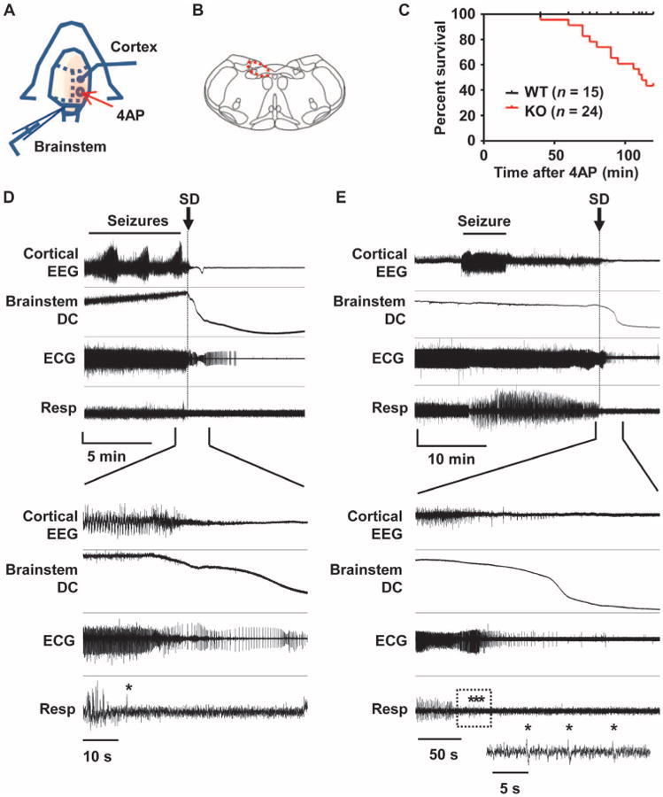
Fig. 1. Premorbid cardiorespiratory dysregulation and brainstem SD in Kv1.1 mutant associated with cortical seizures in vivo.

(A) Diagram of experimental setup for application of 4AP and recording of EEG and brainstem DC potentials in spontaneously breathing urethane-anesthetized juvenile mice (P18 to P25). (B) Illustration of brainstem recording area (red circle). (C) Time until death in Kv1.1 wild-type (WT) and KO mice after focal 4AP application. (D and E) Representative traces of premorbid sequence of the cortical EEG, brainstem DC current, ECG, and respiration in two Kv1.1 KO mice. Expanded traces shown in the lower half of the panels illustrate the temporal association between loss of cortical EEG activity, brainstem SD, and development of cardiorespiratory arrhythmias. Asterisk, gasping. (D) Immediate postictal EEG flattening tightly coupled to onset of cardiorespiratory dysregulation and brainstem SD. Vertical scale: cortical EEG, 0.35 mV; brainstem DC, 5 mV; ECG, 0.22 mV; respiration, arbitrary units. (E) Delayed cortical suppression and cardiorespiratory shutdown >10 min after final intense seizure activity. The respiratory trace in the box is further expanded and shown in the inset. Vertical scale: cortical EEG, 0.31 mV; brainstem DC, 18 mV; ECG, 0.43 mV; respiration, arbitrary units.
