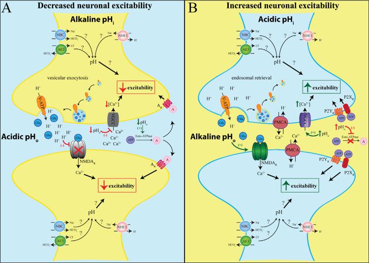
Fig. 1.
The influence of pH on neuronal excitability. When extracellular pH (pHo) is acidic and intracellular pH (pHi) is alkaline, neuronal excitability is decreased. Upon neuronal stimulation, vesicular release of neurotransmitters, such as glutamate (Glu, blue), is accompanied by the release of co-packaged H+. Transient incorporation of the V-ATPase into the plasma membrane induces further expulsion of H+ from the cytosol into the synaptic cleft and causes acidic pHo. The extracellular H+ blocks the voltage-gated Ca2+ channel (VGCC) and reduces presynaptic Ca2+ accumulation, thus inhibiting additional neurotransmitter release and neuronal excitability. Furthermore, acidic pHo also reduces the opening frequency of postsynaptic NMDA receptors (NMDAR), thus decreasing postsynaptic excitability. Extracellular ATP (purple) is broken down into adenosine (pink) driven by ecto-ATPase which is stimulated by low pHo. Adenosine can bind and activate adenosine receptors (AR) and further decrease neuronal excitability. Neuronal excitability is increased when pHi is acidic and pHo is alkaline. The V-ATPase is retrieved from the membrane into early recycling endosomes after neuronal firing, reducing H+ extrusion and elevating pHo. In response to accumulated intracellular Ca2+, the pre- and postsynapse plasma membrane Ca2+/H+ ATPases (PMCA) expel Ca2+ and import H+, further elevating pHo. The alkaline pHo enhances NMDAR activity, facilitating postsynaptic depolarization. Furthermore, alkaline pHo inhibits ecto-ATPases and prevents ATP degradation. The increased availability of ATP activates P2X and P2Y receptors, initiating cascades that could enhance neuronal excitability. Many other pH regulatory players discussed in Section 4 (NHEs, AEs, NBCs) may also contribute to neuronal excitability.

