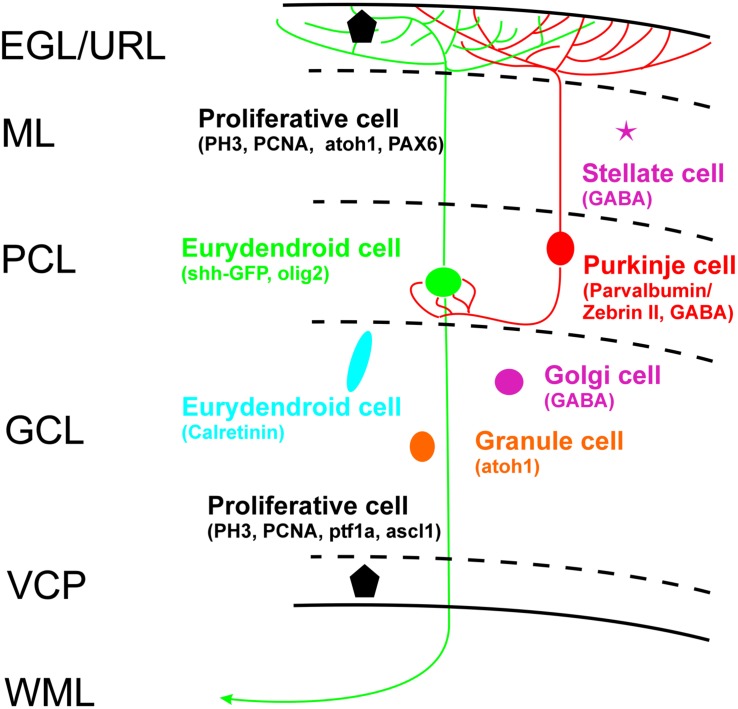
FIGURE 10.
Schema of zebrafish larval cerebellar plate organization summarizes cell types with characteristic cellular markers shown in the present study, with some additional data from Wullimann and Knipp (2000: PCNA), Wullimann and Rink (2001: PAX6), Mueller and Wullimann (2002: BrDU), Wullimann and Mueller (2002: ascl1a), Mueller et al. (2006: GABA), Kani et al. (2010: ptf1a/atoh1a-c), Wullimann et al. (2011: atoh1a-c). EGL, external granular layer; GCL, granule cell layer; ML, molecular layer; PCL Purkinje cell layer; VCP, ventral (ventricular) cerebellar proliferation zone; WML, white matter layer.

