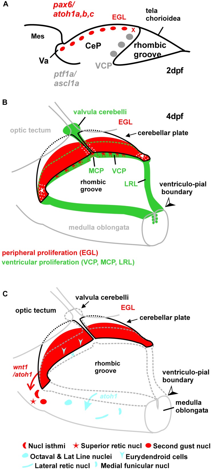
FIGURE 11.
Schematic drawings of zebrafish rhombic lip and cerebellar plate development. (A) Sagittal section showing early larval zebrafish cerebellum (redrawn after Kani et al., 2010). The x indicates site of ventricular upper rhombic lip origin of subpial atoh1 positive cells (see Wullimann et al., 2011). (B) Larval zebrafish rhombic lip (incl. cerebellar) proliferation zones. (C) Migration dynamics involving rhombic lip and external granular layer. The wnt1 derived cholinergic nuclei in the isthmic region were demonstrated in zebrafish larvae (Volkmann et al., 2010). The atoh1a derived structures were observed in adult zebrafish transgenic atoh1a-GFP line (Wullimann et al., 2011). Note that the eurydendroid cells represent only the small atoh1a derived fraction. See text for details. CeP, cerebellar plate; EGL, external granular (or germinative) layer; LRL, lower rhombic lip; MCP, medial cerebellar proliferation zone; Mes, mesencephalon; MO, medulla oblongata; VCP, ventral cerebellar proliferation zone (ventricular germinal matrix); Va, valvula cerebelli.

