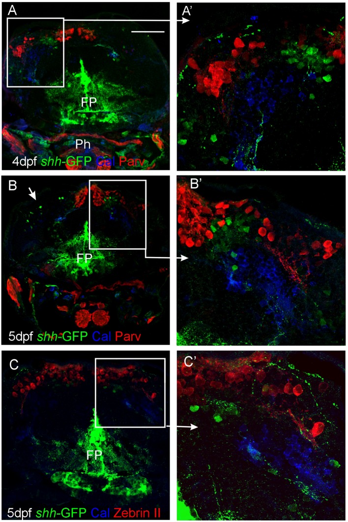
FIGURE 5.
Confocal photomicrographs (optical sections) of transverse sections of shh-GFP zebrafish cerebellum at 4 and 5 dpf immunostained for calretinin/parvalbumin (A,B) or calretinin/Zebrin II (C) with corresponding enlargements to show cellular details (A’–C’). Arrows point out GFP-expressing cells. Scale bar: 100 μm (applies to B and C). FP, floor plate; Ph, pharynx.

