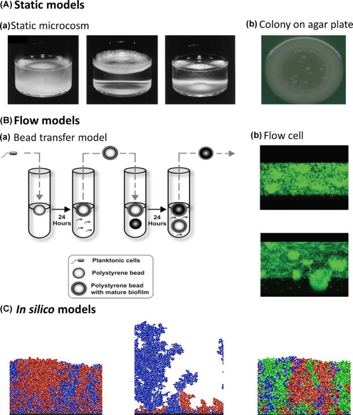
Figure 1.

Illustration of biofilm models. (A) ‘Static models’: (a) The static microcosm model consists of a non-shaking test tube in which a biofilm mat like structure forms at the air–liquid interphase (Adapted from Rainey and Travisano 1998, also used by e.g. Kassen and Rainey 2004; Fukami et al. 2007; Kassen 2009; Spiers 2014). (b) Colonies on agar plates are considered to be suitable biofilm models due to the presence of gradients, an increased mutation rate and a structured environment (Adapted from Kim et al. 2014, also used by e.g. Korona et al. 1994; Perfeito et al. 2008; Koch et al. 2014; Saint-Ruf et al. 2014; van Gestel et al. 2014). (B) ‘Flow models’: (a) In the bead transfer model, plastic beads are put into slowly rotating test tubes. Biofilms grow on the beads and experience flow conditions due to the rotation of the test tubes. In every transfer cycle, the colonized beads are put in a new tube with new beads, without adding new bacteria (Adapted from Poltak and Cooper 2011, also used by e.g. Poltak and Cooper 2011; Traverse et al. 2012; Ellis et al. 2015; O'Rourke et al. 2015). (b) In flow cells, biofilms can grow in the presence of unlimited nutrients and dispersion (Adapted from Kirisits et al. 2005, also used by e.g. Boles, Thoendel and Singh 2004; Hansen et al. 2007; Koh et al. 2007; Yarwood et al. 2007; Lujan et al. 2011; Tyerman et al. 2013; McElroy et al. 2014; Penterman et al. 2014; Udall et al. 2015). (C) ‘In silico models’: agent-based in silico models consist of single dividing cells (the agents) that are programed to grow until a certain radius and then divide. Different parameters can be easily included and adapted (Adapted from Mitri, Xavier and Foster 2011, also used by e.g. Xavier and Foster 2007; Nadell et al. 2008; Nadell, Foster and Xavier 2010; Kim et al. 2014; van Gestel et al. 2014; Schluter et al. 2015).
