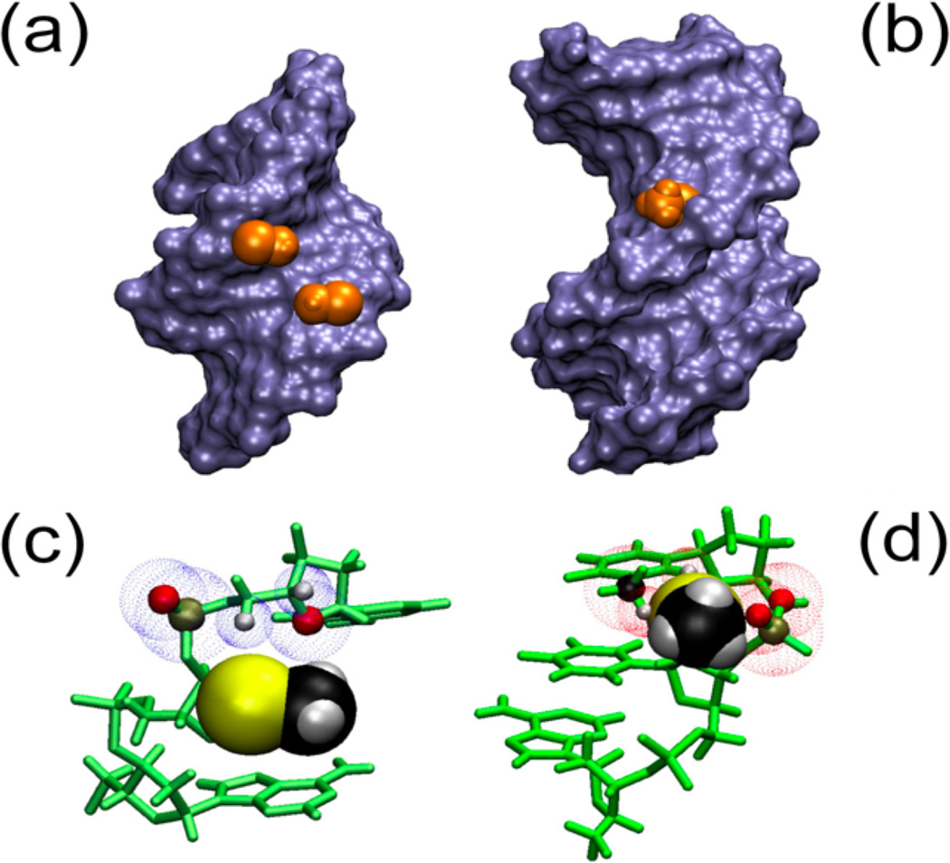
Figure 6.
2’-SeCH3 groups implanted in A and B helical structures. Panels A and B represent Connelly surfaces with the Se-CH3 group depicted as Van der Waals (VdW) representations in orange. In panel C and D the bases and sugars are shown in green and Se-CH3 is depicted as VdW spheres. A) The group is nestled comfortably in the minor groove of an A-type helix (pdb: 1MA8). C) No clashes are apparent between the 3’ neighboring residue and –SeCH3. The blue dotted spheres depict VdW spheres of close atoms. B) In a B-type helical model (dGCGAAUSeMeTCGC), the Se-CH3 group points away from the major groove but experiences significant clashes with the backbone as well as the base on the 3’ side of the modification, which would disrupt base stacking. D) Predicted clashes for the B helix model are indicated with red VdW spheres for backbone (O and P) as well as the base (CH3 and H6).

