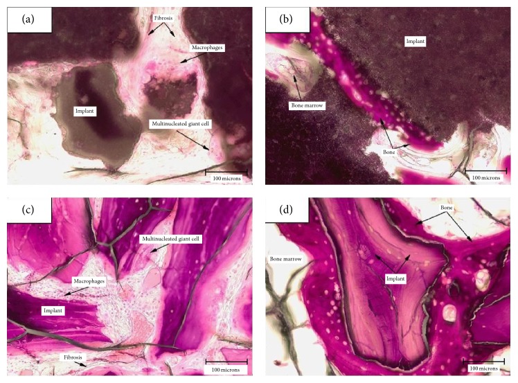
Figure 4.
Microscopic evaluation after 12 weeks (20x objective). (a) Inflammatory cells and fibrosis surrounding the implant (SB) in the cortical area of the implant; (b) new bone has formed along the surface of the implant (SB) located in the medullary area of the defect without inflammation; (c) inflammatory cells and fibrosis surrounding the implant (BS) in the cortical area of the implant; (d) new bone has formed along the surface of the implant (BS) located in the medullary area of the defect without inflammation.

