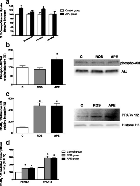
Fig. 6.

Analysis of APE effects on the signaling pathways involved in GLUT4 translocation in L6 myotubes. a Cells were pre-incubated with inhibitors of PI3K (100 nmol/L wortmanin), ERK1/2 (30 μmol/L PD98059) and PPARγ (10 μmol/L GW9662) and then incubated in the absence or presence of 25 μg/mL APE for 2 h. Next, 2-DG uptake was determined. b, c L6 myotubes were deprived of FBS for 18 h and then incubated with 10 μmol/L rosiglitazone (ROS) or 25 μg/mL APE for 30 min. Akt phosphorylation b in cell lysates, and PPARγ levels c in nuclear extracts were measured. d CHO-k1 cells were deprived of FBS for 18 h and then incubated with 10 μmol/L ROS or 25 μg/mL APE for 4 h. PPARγ 1/2 mediated transcription was measured. Results are expressed as mean ± SEM of five independent experiments. (a) p < 0.05 compared with control non-treated cells
