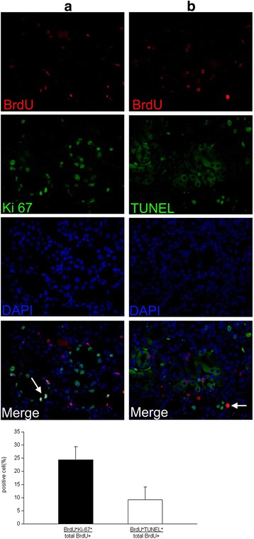
Fig. 8.

Contribution of BrdU+ LRCs in the regeneration of I/R injured mouse kidneys. a Double-immunofluorescence staining for BrdU (red) and Ki67 (green) to visualize the role of BrdU+ LRCs in cell proliferation 4 days after IRI. Newly generated cells formed a special niche-like structure (green), and only some of the BrdU+ LRCs co-expressed Ki-67 in this area (arrows). b Double immunofluorescence was used to determine BrdU+ LRCs undergoing cellular apoptosis on day 4. Scattered BrdU+ LRCs co-expressed TUNEL (arrows) (arrows indicate the double-stained location; magnification × 400). BrdU 5-bromo-2'-deoxyuridine, LRCs label-retaining cells, I/R ischemia/reperfusion
