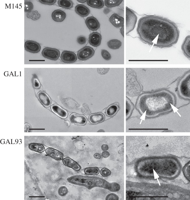
Figure 3.

Transmission electron micrographs of wild-type, sepG mutant and complemented sepG mutant spores. Both a representative overview (left) and close-up (right) are presented. Arrows indicate nucleoids. Note that whereas the wild-type (M145) nucleoid is well condensed and located at the centre of the spores, that of the sepG mutant (GAL1) has an unusual distribution. DNA in the sepG mutant complemented with wild-type sepG (GAL93) was more centrally located than GAL1, although not as well condensed as in wild-type spores. Note the lighter appearance of the spore wall in the sepG mutant. Cultures were grown on SFM agar plates for 5 days at 30°C. Scale bar, 1 µm.
