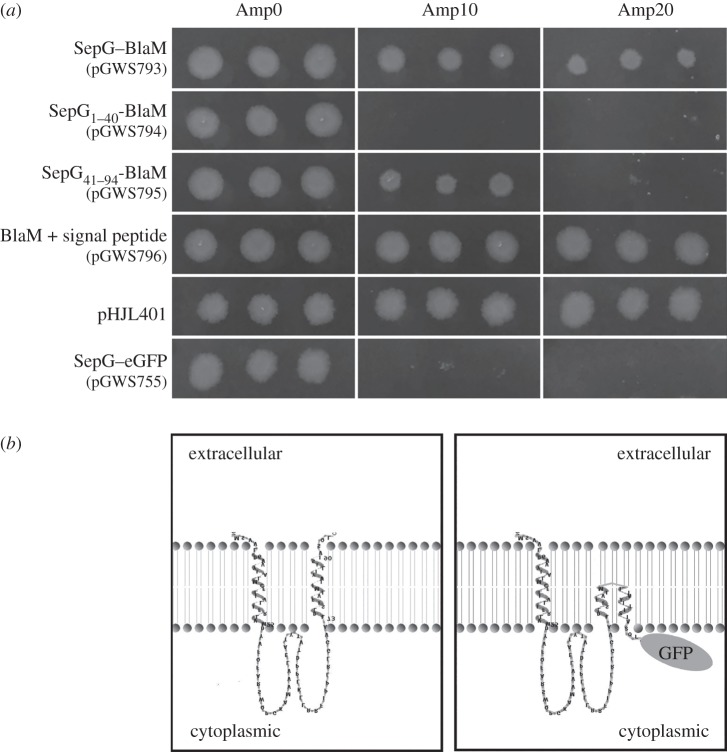
Figure 8.
Membrane topology of SepG. (a) Growth of E. coli JM109 carrying plasmids expressing different BlaM fusions, namely full-length SepG–BlaM (pGWS793); N-terminal SepG1-40-BlaM (pGWS794; first TM domain of SepG); C-terminal SepG41–94-BlaM (pGWS795; second TM domain of SepG); BlaM with signal sequence expressed from the ftsZ promoter(s) (pGWS796; positive control); BlaM with signal sequence expressed from its native promoter (pHJL401; positive control); and pGWS755 expressing SepG–eGFP (pGWS755; negative control). Transformants were grown overnight at 37°C on LB agar plates with 0, 10 or 20 µg ml−1 ampicillin. (b) Topological model of SepG. Left, wild-type SepG, with the topology model generated using TMRPRES2D [47]. Right, model of SepG fused to eGFP, drawn based on the wild-type SepG model.

