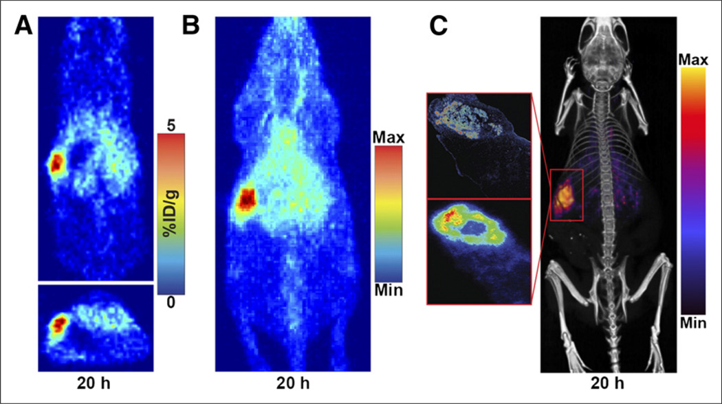
FIGURE 4.
PET and PET/CT imaging of representative mouse with orthotopic Capan-2 xenograft in body of pancreas that was injected with 5B1-TCO followed by 64Cu-NOTA-PEG7-Tz 72 h later. Tomographic slices bisecting tumor (A) and maximum-intensity projection (B) from representative mouse are shown. Also shown is PET/CT scan of same mouse (C, right) and corresponding anti-CA19.9 immunohistochemistry (C, top left) and autoradiography (C, bottom left). %ID/g = percentage injected dose/gram; Max = maximum; Min = minimum.

