
Fig. 2.
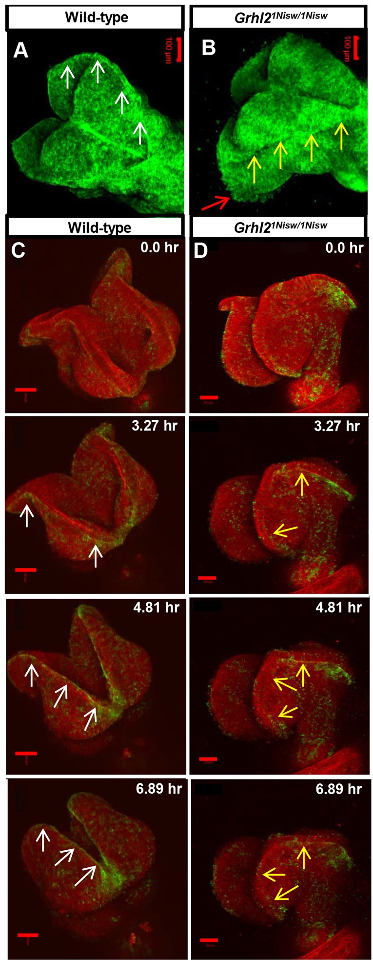
Live imaging reveals dynamic alterations to NNE epithelial integrity in Grhl2 mutants. (A,B) Still images from movies of myr-Venus wild-type and Grhl2 mutant embryos (11 somites; dorsal view). Wild-type embryos exhibit a tight line of membrane association at the neural fold tips (A, arrows) and a smooth external epithelial surface, whereas Grhl2 mutant embryos lack the defined membrane association (B, yellow arrows) and have a rough epithelial surface (red arrow) as the neural folds fall away. (C,D) Time series still images from live imaging of closure point 3 of mTmG:Grhl3Cre/+ wild-type and Grhl2 mutant embryos (11 somites). Wild-type NNE cells exhibit tight membrane association at the NE border (C, arrows) as the folds move toward the midline. Neural folds of Grhl2 mutants fall away and do not meet at the midline, and the membrane association at the NNE/NE border is discontinuous and ill-defined (D, arrows). Scale bars: 100 µm.