
Fig. 3.
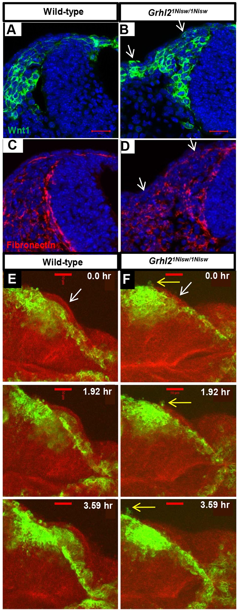
Aberrant localization of migrating NCCs reflects loss of NNE epithelial integrity in Grhl2 mutants. (A-D) Fibronectin immunostaining and NCC visualization (mTmG:Wnt1Cre) shows NCC migration between the NE and NNE and an intact basement membrane under the NNE in wild type (A,C), whereas in Grhl2 mutants the NNE layer contains a few NCCs and this correlates with regions of basement membrane disruption (arrows, B,D). Three embryos per genotype were analyzed. (E,F) Time series from live imaging of mTmG:Wnt1Cre/+ wild-type and Grhl2 mutant embryos (11 somites at start; dorsal view) showing NCCs at the edge of the closing neural folds and below the single layer of NNE (E,F, white arrows). In wild-type embryos, NCCs always move internally (E). In Grhl2 mutants, a few NCCs are seen within the NNE layer and they extend cellular processes externally (F, yellow arrows). Scale bars: 20 µm in A,B; 50 µm in E,F.