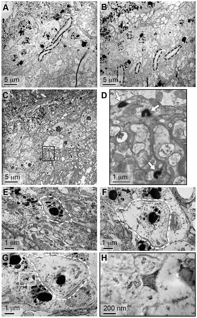
Fig. 5.

Neurodegeneration is associated with laminal deposits. Ultrastructural analyses of lamina in (A) wild-type, (B) dbb1 and (C) bgm1 mutant animals reveal the lamina of mutants to be highly disorganized. Note the parallel organization of photoreceptor axons in wild-type brains, and their misorientation in dbb mutants (outlined in black in A and B). These structures are absent from bgm mutants in which densely stained inclusions are abundant (e.g. boxed region in C). (D) The boxed region from panel C was imaged at higher magnification, and inclusions are indicated with arrows. (E) In wild-type animals, monopolar neurons (outlined in white) are distinguished by their nucleolar content. In bgm dbb double mutants (F,G), however, monopolar neurons are distinguished by cytoplasmic swelling and lytic cell death. Lysing monopolar neurons are oftentimes found in association with extracellular precipitates (arrow in F and boxed in G). (H) The precipitate from panel G was imaged at higher magnification, revealing it to be granular and thus structurally identical to previously described fatty acid precipitates.
