Fig. 2.
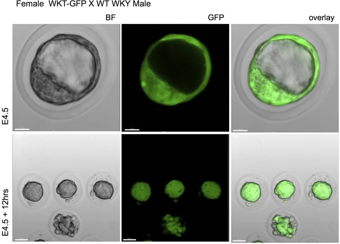
GFP expression in embryos. Female WKT-GFP rats were crossed with wild-type WKY male rats and one-cell embryos removed and imaged under confocal microscope. Representation of three experiments from one-cell embryos at E4.5 and E4.5 plus 12 h showing bright field (BF) images and expression of GFP. Scale bar: 15 μm (top panel); 40 μm (bottom panel).

