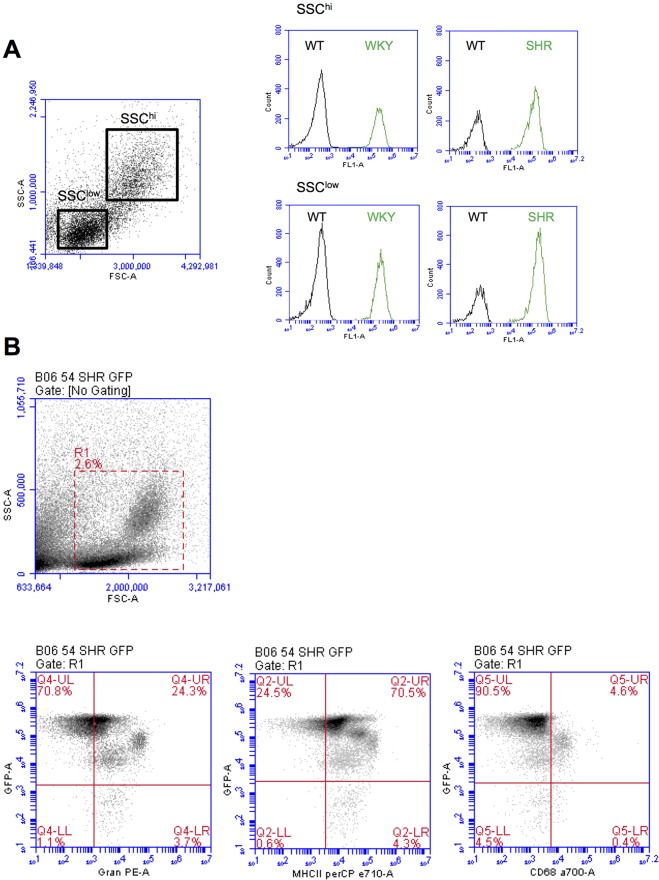
Fig. 4.
GFP expression in blood leukocyte populations. Anticoagulated blood from wild-type, WKY-GFP and SHR-GFP rats were examined for GFP expression with or without fluorescent-labelled antibodies using flow cytometry. (A) Histograms of GFP expression in SSChi and SSClow populations. (B) Lower panels: dot plots of GFP expression versus granulocyte (Gran)-, MHC class II (MHC-II)- or CD68-positive populations, in cells gated by R1 (upper panel). Representative of at least n=4 rats.

