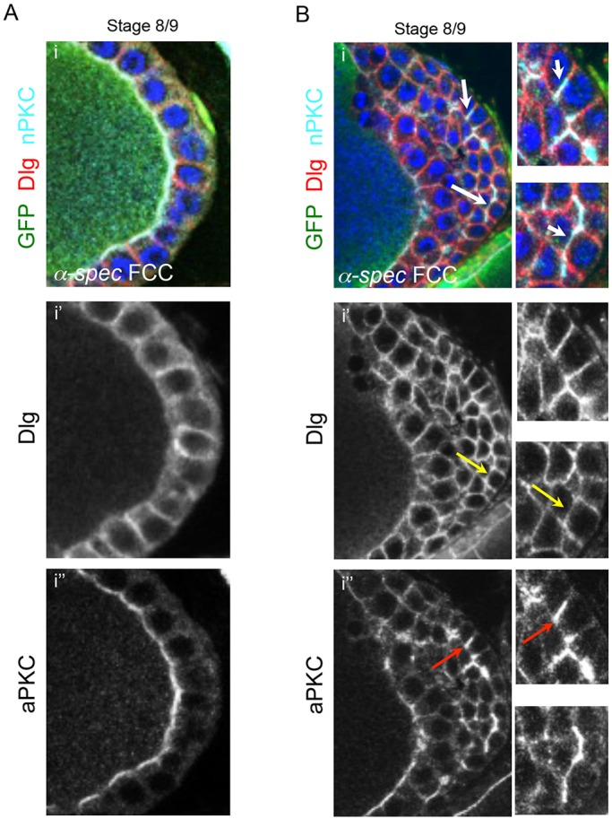
Fig. 2.

α-Spectrin mutant follicle cells display epithelial polarity defects in ectopic layers. (A) The lateral marker Dlg and the apical marker aPKC localize correctly in α-Spec mutant cells that form a monolayer (n=12). (B) In a multilayered α-Spec mutant FE, correct localization of Dlg and aPKC is observed in germline-contacting FCs. In ectopic layers, Dlg is often mislocalized between ectopic layers of cells (i′, yellow arrows), and aPKC often expands into the lateral membrane (i″, red arrows). The white arrows in the top panel indicate colocalization between the mislocalized markers. Some degree of polarity is still preserved because Dlg and aPKC are generally localized correctly in α-Spec ectopic layers (n=35). All images show the posterior domain of S8/9 egg chambers. Blue, DAPI; red, Dlg; light blue, aPKC. α-Spec mutant clones (FCCs) lack GFP.
