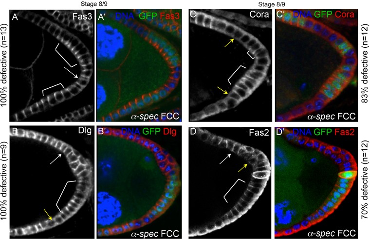
Fig. 5.
The apicolateral localization of Fasciclin 3, Discs large, Coracle and Fasciclin 2 is lost in α-Spectrin mutant cells. (A-D′) At S8/9, the apicolateral localization of the SJ proteins is lost in α-Spec mutant FCs (α-Spec FCCs) that form a monolayered FE (arrows), compared with their neighboring control FCs (brackets). The proteins sometimes extend along the entire lateral membrane (white arrows) or are absent altogether (yellow arrows). The percentages indicate the ratio of egg chambers containing partial posterior clones in a monolayer that display this defect. The number of mutant cells in each egg chamber was at least five. All merged images show DAPI in blue, and Fas3, Dlg, Cora or Fas2 in red. α-Spec FCCs lack GFP.

