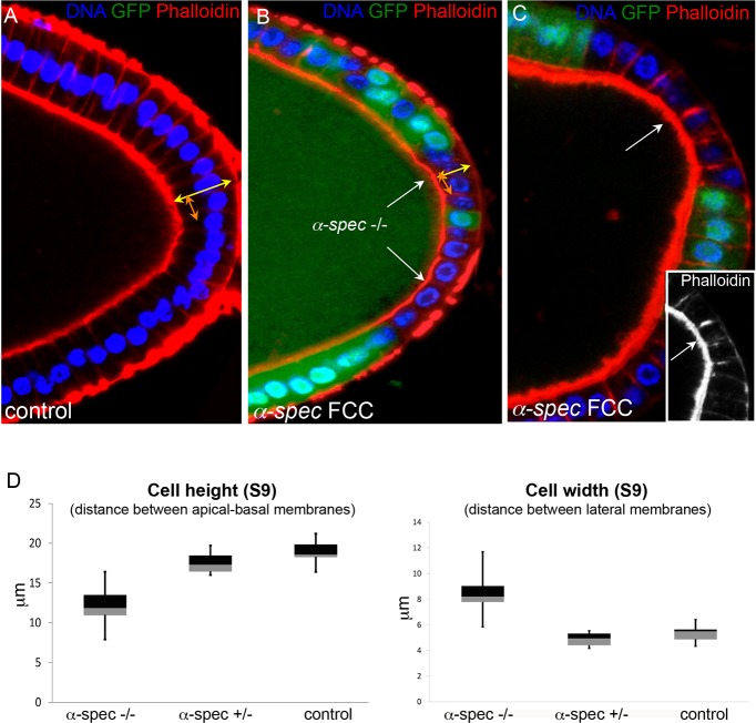
Fig. 6.
α-Spectrin mutant follicle cells fail to form a columnar epithelium. (A) Most FCs expand their lateral membrane at mid-oogenesis, changing shape from cuboidal to columnar (see also Figs S1 and S4). (B,C) Contrary to wild-type FCs, α-Spec cells (α-Spec FCCs, α-Spec−/−) do not extend properly their lateral membrane and fail to become columnar. (C) F-actin levels appear to be higher in α-Spec mutant cells. A-C show posterior FCs. DAPI in blue, Phalloidin in red. All α-Spec cells lack GFP. White arrows indicate the mutant clone. (D) Box plot quantification at S9. The mean height (yellow arrows in A,B) of wild-type and α-Spec cells is 18.95 and 12 µm, respectively. The mean width (orange arrows in A,B) of wild-type and α-Spec cells is 5.30 and 8.44 µm, respectively. The Welch two-tailed t-test P-values between wild type and α-Spec mutant (−/−) for cell height and width are 1.123×10−06 and 0.0002, respectively. Measurements were performed in posterior cells that maintain a monolayer. n=40 (four cells in ten egg chambers) for all samples.

