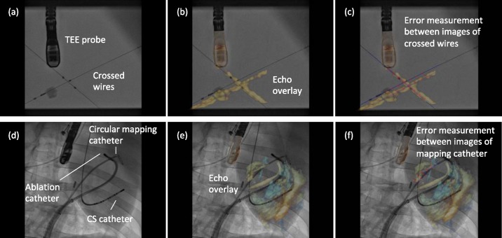
Fig. 3.
(a)–(c) Phantom model experimental overlay of fluoroscopic and echocardiographic images. Errors were measured between automatically defined points on straight line models of the crossed wires. (d)–(f) Porcine experimental study overlay. Errors are measured as the shortest distance from automatically defined points on the echo catheter image to a spline model of the X-ray catheter.

