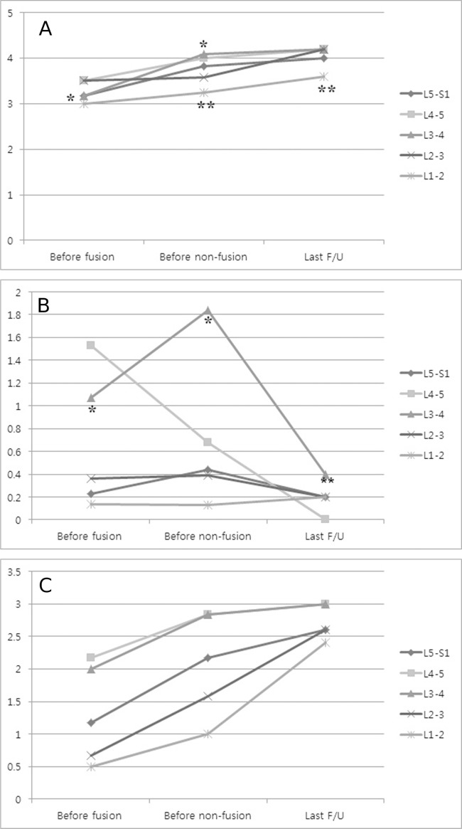
Fig. 4.
MRI changes. A) Disc degeneration. Each segmental lumbar intervertebral disc gradually degenerated from the time before fusion surgery to after non-fusion surgery. Before non-fusion surgery, disc degeneration at L3-4 (*) showed statistically significant changes compared to that before fusion surgery (p=0.046). Between non-fusion surgery and the last MRI evaluation, the change at L1-2 (**) was statistically significant (p=0.032). B) Central stenosis. In the state between fusion and non-fusion surgery, stenosis at L3-4 (*) demonstrated significant degeneration (p=0.041) despite the fact that the L4-5 segment was decompressed by the fusion surgery. After non-fusion surgery, L3-4 (**) sufficient decompression was accomplished (p=0.041), but other instances of segmental stenosis did not show statistically significant changes. C) Facet joint degeneration. Each segmental facet joint was degenerated as time passed. However, the changes between fusion and non-fusion surgery and between non-fusion surgery and the last follow-up did not show statistical significance.

