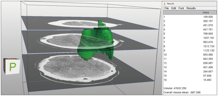
FIGURE 1.
Volumetric analysis of pneumocephalus was calculat-ed using ImageJ. The pneumoce-phalus of subdural space on axial brain computed tomography was bounded as region of interest which multiplied by the slice thickness. And all slice volumes were added up to estimate the vo-lume of each 3D structure.

