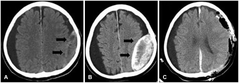
FIGURE 1.
A: Incompletely resolved chronic subdural hematoma after burr hole trephination 4 years prior to admission. A thick-walled isodense lesion is seen in the left temporoparietal cerebral convexity. B: Preoperative computed tomography scan shows a lentiform lesion (8×3.8 cm) with high density partially mixed with isodensity to low density, in the left temporoparietal cerebral convexity. Large amounts of subdural fluid collection along both cerebral convexities are seen. C: Postoperative CT scan shows a newly developed hematoma. An acute subdural hematoma along the interhemispheric fissure and left cerebral convexity is seen.

