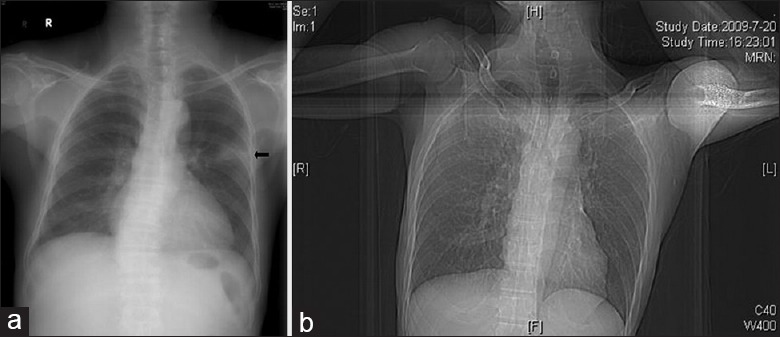
Figure 2.

Representative chest X-rays of human immunodeficiency virus-positive and human immunodeficiency virus-negative patients with Talaromyces marneffei infection. (a) Male human immunodeficiency virus (negative), 71-year-old with chronic lymphocytic leukemia, the X-ray shows patchy shadows in the upper lobe of the left lung (arrow). (b) Male, human immunodeficiency virus (positive), 54-year-old with repeated fever for 2 weeks, the X-ray shows nearly normal in both lungs.
