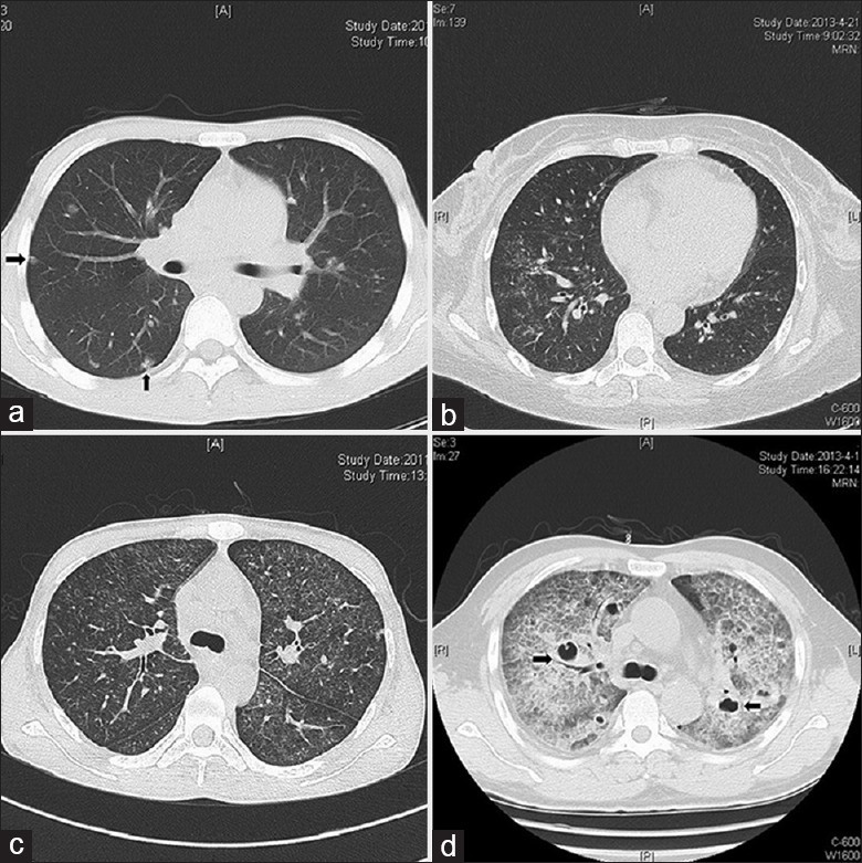
Figure 3.

Representative computed tomography images of human immunodeficiency virus-positive and human immunodeficiency virus-negative patients with Talaromyces marneffei infections. (a) Male, 31 years, human immunodeficiency virus (positive), computed tomography shows multinodular lesions (arrows) in both lungs. (b) Female, 47 years, human immunodeficiency virus (positive), computed tomography shows scattered patchy shadows in both lungs. (c) Male, 31 years, human immunodeficiency virus (positive), computed tomography shows diffuse ground-glass-like lesions in both lungs. (d) Male, 56 years, human immunodeficiency virus (negative), computed tomography shows diffuse interstitial lesions with cavity-like changes (arrows) in both lungs.
