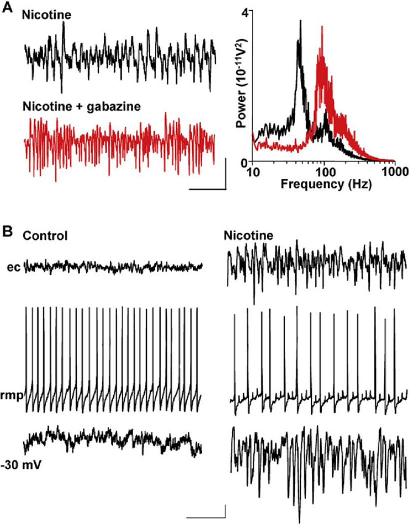
Figure 2. GABAA Receptor Activation Is Critical for Gamma Generation but Not VFO Generation.
(A) Gabazine (1 µM) abolishes gamma rhythms but enhances VFO in field recordings. Example traces are field recordings from the Purkinje cell layer. Note the attenuation of gamma frequency activity and the predominance of VFO activity on blockade of GABAA receptors. Spectrograms are taken from 60 s epochs of field data (n = 5): black line, data in the presence of nicotine alone; red line, nicotine and gabazine. Scale bars: 0.1 mV, 100 ms.
(B) Purkinje cell responses to nicotine application show gamma frequency inhibitory postsynaptic potentials. When bathed in aCSF alone, no rhythmic activity was seen in field potential recordings, despite continuous action-potential generation in individual Purkinje cells (left panel). The Purkinje cell shown has a mean rmp of 58 mV. When depolarized to 30 mV by current injection, only very few, small IPSPs were seen. Scale bars: 50 µV (field), 10 mV (rmp), 2 mV (30 mV), 100 ms. In the presence of nicotine (right panel), Purkinje cell layer field potentials showed high-frequency oscillatory activity and individual Purkinje cells hyperpolarized by 4 mV. Purkinje cell spike rates were approximately halved, and multiple partial spikes (spikelets) were seen. Mean membrane potential of the Purkinje cell shown was 62 mV. Depolarization to 30 mV exposed intense trains of IPSPs at gamma frequency, phase locked to the field potential rhythm. Scale bars are as in the left panel.

