Fig. 1.
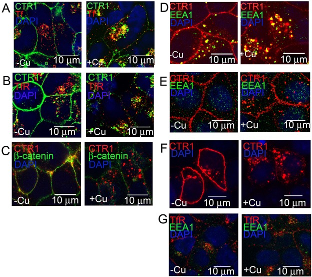
CTR1 co-localization with membrane or endosome markers after Cu treatment. (A) HEK-CLIP–CTR1 cells were incubated with CLIP-Surface–488 (green), followed by treatment with or without 30 µM Cu and with fluorescent transferrin (red) (30 min), and live cell imaging was performed. (B–D) For fixed cell immunofluorescence, HEK-FLAG–CTR1 cells were treated with or without Cu, fixed cells were labeled with anti-FLAG for CTR1 and either anti-TfR (B), anti-β-catenin (C), or antibody against early endosome marker EEA1 (D). (E) MDCK-FLAG–CTR1 cells treated with or without Cu were double-labeled with anti-FLAG for CTR1 and anti-EEA1. (F) SKCO15 cells transiently transfected to express FLAG-tagged CTR1 were treated with or without Cu (30 min), fixed, probed with anti-FLAG (CTR1). (G) HEK-CLIP–CTR1 cells treated with or without 30 µM Cu (30 min), fixed, and labeled with anti-TfR, anti-EEA1 and DAPI. Stained nuclei are visible in blue and merged fluorescence is displayed in yellow. Scale bars: 10 µm.

