Fig. 3.

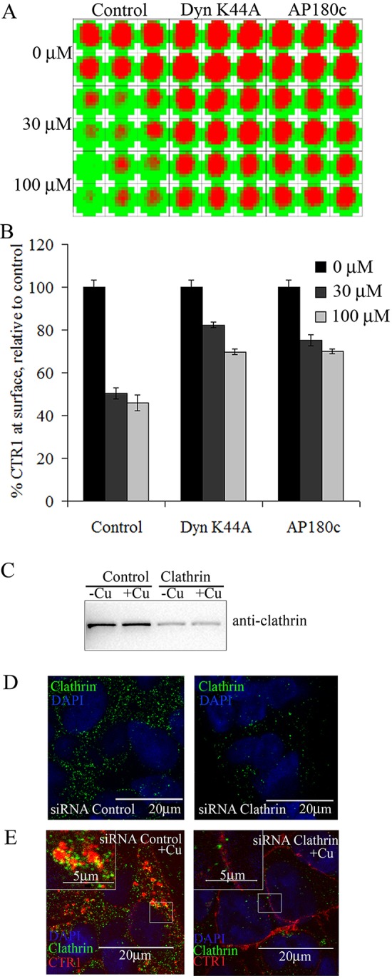
Cu-induced CTR1 internalization is mediated by clathrin and dynamin pathways: plate assay. HEK-CLIP–CTR1 cells transfected with empty vector control, DynK44A, or AP180c were treated with 30 or 100 µM Cu for 30 min, and surface CTR1 was labeled with fluorescent CLIP-Surface–547. (A) An example of a plate scan from the CLIP plate assay using a 10×10 scan matrix for each well. Red indicates CLIP fluorescence on surface CTR1, and green indicates lack of fluorescence. (B) Histogram showing the relative surface expression of CTR1 after Cu treatment compared with untreated control cells, mean±s.e.m., n=6. (C–E) HEK-CLIP–CTR1 cells were transiently transfected with siRNA against either scramble control or clathrin-targeted siRNA. (C) Lysates from siRNA-transfected cells treated with or without excess Cu were collected and subject to western blot analysis using anti-clathrin antibodies. (D) Cells were probed using anti-clathrin antibodies to show sufficient knockdown of clathrin (right panel) compared with control (left panel). (E) siRNA-transfected cells were labeled with Surface-CLIP (red) and then treated with or without 30 µM Cu for 30 min prior to fixation and anti-clathrin (green) immunofluorescent probing. Zoomed insets of selected regions are displayed. Scale bars: 20 µm, 5 µm in insets.
