Fig. 4.

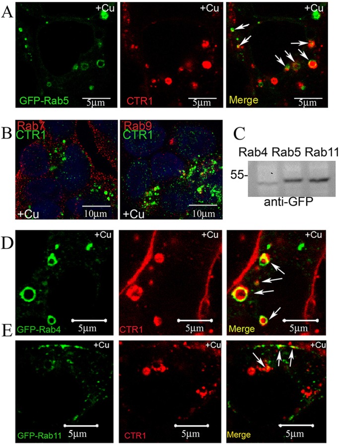
Internalized CTR1 co-localizes with early endosomes, but not with late endosomes. (A) HEK-CLIP–CTR1 cells expressing GFP–Rab5 were labeled with CLIP-Surface–547, treated with 30 μM Cu for 30 min, and subject to confocal microscopy. Intracellular compartments containing co-localized Rab5/CTR1 are visible in yellow (arrows). (B) HEK-FLAG–CTR1 cells treated with 30 μM Cu for 30 min were fixed and double-labeled with anti-FLAG (CTR1) and either anti-Rab7 or anti-Rab9 antibodies. (C) HEK-CLIP–CTR1 cells were transiently transfected with plasmids to express either GFP–Rab4, GFP–Rab5, or GFP–Rab11. Expression in total cell lysates was determined by western blot. (D,E) For live cell confocal fluorescence, transfected cells were labeled with CLIP-Surface and treated with 30 μM Cu for 30 min. CTR1 is internalized from the plasma membrane during Cu treatment and then co-localizes (arrows) with Rab4 (D) and Rab11 (E) GTPases. Scale bars: 5 μm in A,D,E; 10 μm in B.
