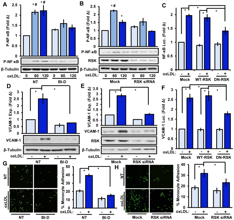
Fig. 3.
RSK mediates oxLDL-induced NF-κB activation, VCAM-1 expression and monocyte adhesion. (A,B) Endothelial RSK signaling was inhibited with (A) BI-D1870 (BI-D, 5 μM, 1 h) or (B) RSK1 and RSK2 siRNA, and oxLDL (100 μg/ml)-induced NF-κB phosphorylation (P-NF-κB) was determined by immunoblotting after 60 or 120 min (n=4). (C) BAE cells were co-transfected with an SSRE-luciferase construct and either wild-type (WT)-RSK or dominant-negative (DN)-RSK constructs. OxLDL-induced luciferase (Luc.) activity was determined after 24 h (n=3). (D,E) RSK signaling in HAE cells was inhibited with (D) BI-D1870 (5 μM, 1 h) or (E) RSK1 and RSK2 siRNA and oxLDL-induced VCAM-1 expression was determined after 6 h by immunoblotting (n=4). (F) BAE cells were co-transfected with a VCAM-luciferase construct and either WT-RSK or DN-RSK constructs. OxLDL-induced luciferase (Luc.) activity was determined after 24 h (n=3). (G,H) RSK signaling in HAE cells was inhibited with (G) BI-D1870 (5 μM, 1 h) or (H) RSK1 and RSK2 siRNA, and oxLDL-induced monocyte adhesion was determined after 6 h using static adhesion assays (n=4). Results are mean±s.e.m. *P<0.05 (compared to 0 time or no treatment) or #P<0.05 (compared to respective timepoint) using two-way ANOVA with Bonferroni post-hoc test. NT, no treatment. Mock indicates mock transfection.

