Fig. 5.
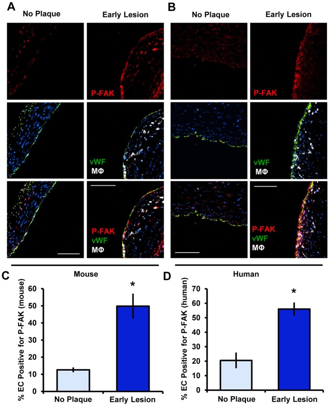
FAK activation in endothelial cells is detected during early atherosclerosis. (A) Mouse lesions were immunostained for active FAK (P-Y397 FAK, red), endothelial cells (vWF, green), myeloid cells (Mac2; MΦ, white) and DAPI (blue) (n=4 for each group). (B) Human coronary arteries were immunostained for active FAK (P-FAK, red), endothelial cells (vWF, green), myeloid cells (CD68, MΦ, white) and DAPI (blue) (n=4 for each group). (C,D) Endothelial FAK phosphorylation in the mouse and human specimens was quantified as the percentage of the endothelial cell layer (EC, vWF positive) staining positive for P-FAK. Results are mean±s.e.m. *P<0.05 (compared to no plaque result) using two-tailed Student's t-test. Scale bars: 50 μm (A); 100 μm (B).

