Fig. 6.
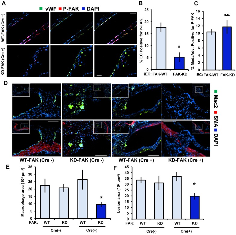
FAK activation in endothelial cells is required for leukocyte recruitment in early atherosclerosis. (A) Aortic roots from FAK-WT (Cre+) and FAK-KD (Cre+) transgenic mice were immunostained for P-Y397 FAK (red), an endothelial cell marker (vWF, green) and DAPI. (B,C) P-FAK staining was quantified as the percentage of the endothelial cell layer (EC, vWF-positive) or the vessel media and adventitia staining positive for P-FAK. (D) Aortic root from transgenic mice was immunostained for myeloid cells (Mac2, green), smooth muscle cells (SMA, red) and DAPI. Representative micrographs are shown. (E) The Mac2-positive area was quantified from an average of three separate sites along the aortic root 50 μM away from each other. (F) Lesion size was quantified from neointima formation as an average of three separate sites along the aortic root 50 μM away from each other [n=6 for WT-FAK (Cre−), 5 for WT-FAK (Cre+), 7 for KD-FAK (Cre−) and 5 for KD-FAK (Cre+)]. Results are mean±s.e.m. *P<0.05 (compared to FAK-WT) using two-tailed Student's t-test. Scale bars: 20 μm (A); 50 μm (D).

