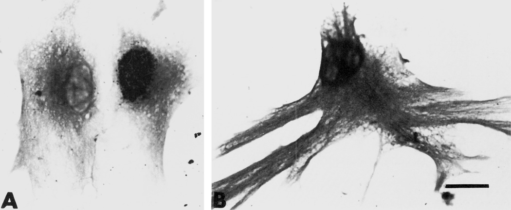
Figure 6.
(A and B). Brightfield photomicrographs of glial fibrillary acidic protein (GFAP)-immunoreactive astrocytes. Compared to astrocytes treated with growth media alone (A, controls); many of those treated with 10−6 M morphine show a greatly exaggerated increase in both size and the elaboration of cytoplasmic processes (B). Scale bar = 25 µm.

