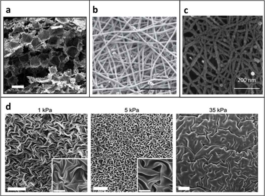
Figure 3. Synthetic and semi-synthetic biomaterials.

Scanning electron micrographs of (a) porous poly-lactic-glycolic scaffolds; reproduced with permission from [37] and (b) electrospun polycaprolactone scaffold; scale bar = 200 µm; reproduced with permission from [39]. (c) Scanning electron micrograph of a self-assembling peptide hydrogel; reproduced with permission from [49]. (d) Scanning electron micrograph of dehydrated hyaluronic acid gels; scale bar = 20 µm; inset scale bar = 5 µm; reproduced with permission from [45].
